 +44 (0)1582 519755
 enquiries@lawsonscientific.co.uk
contact us
t. +44 (0)1582 519755
e. enquiries@lawsonscientific.co.uk

Get in touch for a quote or to find out more about how we can support you ….
XMT is an ideal non-destructive method for imaging the internal and external micro-structure of your materials.
It utilises the same principles as CT scanners found in hospitals but at a far greater resolution. X-rays are used to image the relative density distribution of ‘slices’ of an object.
These slices are then reconstructed to give a 3D image containing details of the internal structure.
XMT is a particularly valuable technique when analysing drug dissolution, as it provides an image of the pores and channels through which water flows to break down the structure of a tablet.
Lawson Scientific has scientists trained on a Phoenix v|tomo|x Compac, which has a sub-micron maximum pixel resolution.
Lawson Scientific offer a range of microscopy techniques, some of which can be performed wet as well as dry for particles in suspension. Our scientists are well-equipped to analyse most samples and can help you choose the best technique for your requirements.
We have the following microscopy techniques to offer our customers:
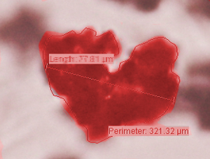
Lawson Scientific have a number of light microscopes in use for particle image analysis.
We have software which allows us to capture, clarify and measure magnified particles according to your requirements.
SEM is often preferred over light microscopy for imaging particles as it provides a higher magnification, better depth of field and images are more 3D. If nonconductive, samples are coated in a conductive material such as gold and placed in a vaccum where an electron beam is systematically traced over it. Dislodged secondary electrons from the surface of the sample are registered on a number of detectors and dot by dot, row by row, an image is built up on a monitor.
SEM is frequently paired with EDX analysis to characterise chemical composition.
SEM is an excellent technique for judging the uniformity of your samples, for example in industries utilising metal powders to cut materials, coatings or to create alloys.
Lawson Scientific have the expertise to help interpret images and select the best imaging methods for your samples.
Analysis by EDX is used reveal the chemical composition of a material.
As with SEM, an electron beam isused to blast a sample, causing the release of electrons from the elements comprising a sample. These electrons leave behind a gap in the inner shell of an element, which is filled by an electron from the outer shell of either the same electron or a neighbouring one. As an electron decreases shell-levels, x-rays are released. The difference in shell-levels between the two electrons determines the number and strength of these x-rays, which are detected and used to discern the chemical composition of a sample.
Often coupled with SEM analysis, Lawson Scientific are able to offer EDX analysis as a standalone procedure or as an add-on with SEM.
As with SEM, TEM works by bombarding a sample, which must be electron transparent, under vacuum with high energy electrons from a gun.
In a similar way to a shadow being created when light is blocked, the number of these electrons detected on the plate below the sample depends on the level of transparency of the sample. TEM is thus able to generate a black and white image at a high resolution that can give detailed images of shape, texture and size down to a molecular level.
TEM is particularly useful to technology companies looking to identify flaws in micro-sized materials. Lawson Scientific has scientists trained on Equipment name who are experienced in TEM and other microscopy techniques, and can image your samples.

t. +44 (0)1582 519755
e. enquiries@lawsonscientific.co.uk
Business Address: Unit 3, Clipstone Brook Industrial Estate, Cherrycourt Way, Leighton Buzzard, LU7 4GP
Registered in England and Wales (Registration Number : 11438299), VAT Number GB 302445934
© 2026 Lawson Scientific
Website by Vantage Agency